Price Vision Group Blog

THE LATEST NEWS FROM PRICE VISION GROUP

15/Jan/2020

Price Vision Group Celebrates 25 Years of LASIK, the first in the state of Indiana.

January 2020 marks the 25-year anniversary of Price Vision Group providing LASIK eye surgery.  We continue to see happy patients from those early years when Dr. Price performed LASIK as part of an investigational study. 
From the start, we were able to correct both nearsightedness and astigmatism. Over time we have continued to refine vision correction options and now provide the most cutting-edge, vision-optimizing treatments available in the U.S.
We began offering refractive surgery in 1983, 37 years ago! We began using a laser to correct vision in 1991. We initially offered surface ablation and in 1995 we began offering LASIK. 
Technological advances in the past 25 years have further improved the safety and accuracy of LASIK. We can now perform topography directed treatments, Contoura™ Vision LASIK, in which we plot 22,000 points on the cornea to generate a topographic map. This allows us to smooth out small imperfections on the corneal surface that cannot be corrected with glasses.  
In the clinical trials that led to FDA approval of topography directed treatments, 30% of the patients could see better postoperatively without any correction than they could see preoperatively using glasses.1 Our patients are achieving these same amazing results.  In fact, most of our patients achieve better than 20/20 vision. 
A vision of 20/20 means that your vision is normal, but surprisingly only 35% of all adults have 20/20 vision without some type of correction, such as glasses. If you have 20/20 vision, you can see at 20 feet what most people see at the same distance. If you are 20/16, you can see at 20 feet what a person with normal vision would need to move up to 16 feet to see, and if you are 20/12, you can see at 20 feet what others would need to move up to 12 feet to see.  These are fantastic, life-changing results!  
At Price Vision Group, we are proud to be at the forefront in researching the newest in laser vision correction technology. We will soon be investigating whether we can further improve upon LASIK technology with even more sophisticated imaging and analysis.  Both the topography-directed treatment and the next-generation treatment rely more on objective imaging of the eye rather than relying so heavily on the subjective assessment of “which is better, 1 or 2”, for greater precision.
As we look back with pride at all that we have accomplished in 25 years, we are also excited about the new technologies we are and will be investigating to provide our patients with the best possible visual outcomes in the safest and most effective ways.


28/Aug/2019

This is an update to a post published on 9/25/17. You can find the original post here.

Descemet’s Stripping Only…a substitute for DMEK?

 By Francis W. Price, Jr., MD & Matthew T. Feng, MD

We are always looking for better ways to correct the visual difficulties of patients with Fuchs’ dystrophy.  In fact, that is why we helped pioneer the evolution of corneal transplants from full-thickness grafts (penetrating keratoplasty) to PLK/DLEK to DSEK to DMEK.  Interest is building now to eliminate the transplant altogether and allow a person’s own endothelial cells to redistribute in a procedure called Descemet’s Stripping Only.
The corneal endothelium is a single layer of cells that lines the back surface of the cornea. These cells pump water out of the cornea to keep it crystal clear. In Fuchs’ dystrophy, these cells become unhealthy and die off, starting in the center and moving toward the periphery over time.  This allows fluid to build up in the cornea causing swelling and hazy vision. Eventually, blisters can develop; this can be painful and cause light sensitivity.

The corneal endothelial cells are attached to a thin membrane called Descemet’s membrane. In Fuchs’ dystrophy, abnormal deposits called “guttae” accumulate on Descemet’s membrane.  These guttae are like water drops on a windshield (guttae mean raindrops in Latin).  They distort the light coming into the cornea and also cause glare and halos and must be removed to improve vision.

A healthy endothelium compared to a diseased endothelium.

DMEK is a tried and true treatment for Fuchs’ dystrophy in which we remove the central corneal endothelium and Descemet’s membrane and implant healthy donor tissue, which has normal endothelial cells and a clear Descemet’s membrane without guttae.  It provides rapid and reliable visual recovery within 2 days to one month after surgery with minimal risk of immunologic graft rejection (<1% with appropriate use of eye drops).[reference] With DMEK, the risk of developing glaucoma is reduced because of the improved corticosteroid eye drop dosing regimens we have developed, and long term graft survival is excellent.
In contrast, we and others have tried Descemet’s Stripping Only (DSO) which is performed in the same manner as with DMEK, except a smaller area of the central area of Descemet’s Membrane is removed and no donor endothelium is transplanted.  We then wait to see if the surrounding endothelial cells migrate from the periphery to recover the central area.  There are two benefits of DSO that excited us. First, there would be no risk of immunologic graft rejection (0% rejection rate) since we are not using a donor cornea so there is no immune response to foreign tissue. Second, the need to use long term topical corticosteroids to prevent graft rejection would be eliminated thereby reducing the side effect of intraocular pressure elevation that can affect up to one-third of transplant recipients.
In a prior analysis of DSO, we removed a 6-mm diameter area of the diseased Descemet’s membrane but we found that recovery was incomplete—the endothelial cells never fully filled in the area of cells that we removed, even after waiting for months.
We, along with other surgeons, are now removing smaller areas of Descemet’s membrane (4 to 5 mm diameter area) and have a higher rate of clearing.  Despite these successes, DSO is not yet a replacement for DMEK for two primary reasons: clearing a smaller area may not always be sufficient to provide the best possible vision, and there is still the potential for prolonged corneal swelling that may last for months hindering visual recovery. By way of comparison, we typically remove unhealthy Descemet’s membrane and endothelium from a much larger area (over twice as big) when performing DSEK or DMEK. However, DSO may be a suitable first-line surgical treatment option prior to DMEK, for those willing to see if stripping alone will satisfactorily resolve their vision problems. If after a few months their cornea is not clear, they would then be a candidate for DMEK.
We have been told that patients are happy with the vision they get using a 4-mm area of treatment. But we wonder if they just notice some visual improvement with the central guttae removed without realizing how much better it could be if a wider area was treated. We know from laser refractive surgery (LASIK or PRK) that a small treatment zone causes patients to have significant glare and halos at night.  We also know that the intraocular lenses used in cataract surgery do not perform well when the clear optical zone is only a 4-mm diameter. Typically, clear zones of 6-mm diameter or more are needed to avoid glare and halos.
Many of our Fuchs’ dystrophy patients tell us they only need one eye treated because their other eye is doing fine.  But after DMEK, they realize the “good” eye was not as good as they thought, and then they want to have the second treated as soon as possible to eliminate glare and haze.  Similarly, in the wintertime when it snows, you can drive your car after just clearing a small area of the windshield, but obviously, it is much better to clear a larger area to view the periphery. Since guttae generally begin forming in the central portion of the cornea, DSO may be a better option for patients earlier in their Fuchs’ dystrophy onset rather than more advanced cases. Your surgeon will determine in you may be a candidate for DSO, or whether DMEK is more fitting. This is an example of when it’s best to seek evaluation as soon as you observe changes in your vision, as you may have more treatment options at that time.

The second concern with DSO is slow healing. Often with stripping alone, it takes several weeks to months for the cornea to clear.  In some cases it takes over 6 months.  There is evidence that when the cornea is continuously edematous (swollen) from non-functioning endothelium for 6 months or longer, permanent changes occur so that when the edema does resolve, the vision does not return to a level as good as it would have been if the cornea had cleared faster. Two centers found that if they waited over 6 months to replace a failed transplant, the visual results were disappointing.4,5  In contrast, we find that when we replace a failed transplant promptly, the visual results are excellent.6  The longer a cornea remains edematous, the more likely it is that vision will permanently deteriorate.
In summary, we look forward to continuing research to assure the cornea will reliably clear within a month or sooner with DSO and are actively conducting studies to optimize this treatment option for Fuchs’ dystrophy patients.  Two small studies found that the use of a “Rock Inhibitor” eye drop available outside the USA seemed to help the cornea clear faster and more reliably by helping the cells migrate and possibly regenerate.7 If you would like to learn more about DSO and determine if you are a candidate for a study, please call (317) 814-2996. Patients who have already had stripping alone and are dissatisfied with their vision or speed of recovery often remain candidates for DMEK and may benefit from evaluation sooner rather than later.    
Looking ahead, cell culture techniques are improving, and someday we may be able to harvest your blood cells and reprogram them to become corneal endothelial cells.  But until we can either do that or use medications to get your peripheral corneal endothelial cells to reliably heal the central cornea in less than a month, we will continue to recommend DMEK as the optimal treatment option for most patients with Fuchs’ dystrophy who are not able to face a potentially long recovery with DSO.
We certainly live in an exciting time with new developments occurring continually around the world. So stay tuned!

References:

  1. Bleyen et al. Spontaneous corneal clearing after Descemet’s stripping. Ophthalmology 2013l120:215.
  2. Arbelaez et al. Long-term follow-up and complications of stripping descemet membrane without placement of graft in eyes with Fuchs endothelial dystrophy. Cornea 2014 33:1295-1299 (our study)
  3. Koenig SB. Planned Descemetorhexis Without Endothelial Keratoplasty in Eyes With Fuchs Corneal Endothelial Dystrophy. Cornea 2015;34:1149-51.
  4. Baydoun et al. Repeat Descemet membrane endothelial keratoplasty after complicated primary Descemet membrane endothelial keratoplasty. Ophthalmology 2015; 122:8-16.
  5. Cirkovic et al. Clinical and ultrastructural characteristics of graft failure in DMEK: 1-year results after repeat DMEK. Cornea 2015; 34:11-7
  6. Price et al. Repeat Descemet Membrane Endothelial Keratoplasty: Secondary Grafts with Early Intervention Are Comparable with Fellow-Eye Primary Grafts. Ophthalmology 2015;122:1639-44.
  7. Moloney et al. Descemetorhexis Without Grafting for Fuchs Endothelial Dystrophy-Supplementation With Topical Ripasudil. Cornea 2017;36:642-648.

16/Jan/2019


Corneal thickness scan

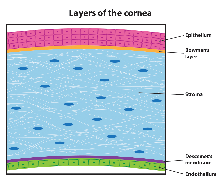
When a patient comes to our office for a LASIK screening, one of the first things we check for is cornea thickness. So why is cornea thickness important for LASIK? During LASIK, an excimer laser is used to remove tissue (ablation) and reshape the cornea so images can focus accurately onto the retina. Since corneal tissue is permanently removed during the LASIK procedure, we want to make sure there is enough tissue remaining to ensure the cornea is structurally safe.


Layers of the cornea


How much corneal tissue is removed during LASIK?


Typically, we will make a LASIK flap that is about 120 microns thick. Our excimer laser will remove approximately 14 to 16 microns of corneal tissue per diopter of treatment. To ensure that the cornea is structurally safe, there should typically be a minimum of 300 microns (μm) remaining before the LASIK flap is repositioned.

The following animation shows a hypothetical LASIK procedure for a patient that has a -4.00 diopter prescription with a cornea thickness of 500 microns (μm).

  • A 120 micron (μm) LASIK flap is created and lifted.
  • 56-64 microns (μm) of corneal tissue are removed during the ablation process.
  • 316-324 microns (μm) of corneal tissue remain before the flap is repositioned.


If it has been determined that your corneas are too thin for LASIK, we have several options to correct your vision and help get you out of glasses and contacts. Options include making a thinner LASIK flap, PRK, or Visian ICL.

To learn more about your options, schedule a free screening here at Price Vision Group, or call us at (317) 814-2933.


20/Dec/2018

Pioneering cornea surgeon Francis W. Price, Jr., MD of Price Vision Group in Indianapolis, IN is scheduled to become the first in the U.S. to perform the revolutionary LIKE procedure. LIKE (LASIK and Intrastromal Keratoplasty) is the next generation in refractive surgery to correct extreme farsightedness in people who cannot be helped by current LASIK or refractive techniques.

Clear, focused vision depends on the cornea and lens, which refract, or bend, incoming light, projecting it precisely on the retina, which sends visual information to the brain. Farsightedness, or hyperopia, is a condition where the eyeball is too short, causing images to project past the retina and affects about 10-20% of the population in the U.S. This makes up-close objects appear extremely out of focus, while far away objects are less out of focus but still not clear. Glasses or contact lenses are the most common treatments. Surgical options have been limited, especially for those with high degrees of farsightedness, because there is no procedure to extend the length of the eye.
Refractive procedures, such as LASIK, have been successful in treating low levels of farsightedness. Currently, higher levels of farsightedness can only be treated with corrective eyewear, cataract surgery, or in patients without cataracts, with a refractive lens exchange where the natural lens in the eye is replaced with an artificial lens (IOL).  These options work well for those in their 40’s or older, but cause younger people, who still have the ability to focus up close, or accommodate, to lose that ability. There is also more risk with having surgery inside the eye, rather than on the surface as with LASIK.
What is LIKE? (LASIK and Intrastromal Keratoplasty)
LASIK and Intrastromal Keratoplasty, or LIKE, offers a cutting-edge surgical treatment for those with farsightedness starting at 3 diopters and up to 10 diopters, a level which has been previously untreatable with LASIK.
LIKE is a two-part procedure which places a portion of a donor cornea into the stromal, or structural, layer of the cornea and upon healing, LASIK is performed. The insertion of the structural donor layer alters the shape of the cornea so that light and images are precisely focused on the retina resulting in excellent, clear vision. This procedure has similarities to a standard LASIK procedure where a flap is created and then lifted to begin treatment. Unlike LASIK, instead of using an excimer laser to remove tissue (ablation), a donor cornea is implanted (to add tissue). After the vision has stabilized, (~2-3 months), the LASIK treatment will be performed on the donor tissue (not the patient’s tissue) to fine tune the vision.
Dr. Francis Price, Jr., who is a world-renowned surgeon and founder of the Cornea Research Foundation of America, has trained more than 600 eye surgeons on advanced surgical techniques, and routinely cares for the most complex eye conditions in patients from around the world. He, along with other Price Vision Group surgeons, have helped pioneer and perform more cornea transplants and specialized procedures than any other clinic in North America. As a result of his excellent track record in pioneering surgical treatments, he was chosen to become the first in the U.S. to perform this ground-breaking new technique.

Terms:

Stroma – the structural part of the cornea, the window of the eye
Keratoplasty – cornea transplant, placing a donated cornea from another person into some one’s cornea


19/Dec/2018

Indianapolis, IN, November 8, 2018 – Price Vision Group is the first practice in Indiana to implant the new FDA-approved Visian Toric ICL from STAAR Surgical. This new implantable collamer lens (ICL) provides a new solution for patients with moderate to high levels of myopia and astigmatism ‒ in one simple procedure. Previously, ICL patients with astigmatism would need a separate LASIK procedure to correct astigmatism.

Matt, a resident from Lafayette, will become the first patient in Indiana to receive the Visian Toric ICL. “I’ve been in glasses since kindergarten and hard contacts since fifth grade… so I’m excited,” stated Matt just before his surgery. The procedure to insert the Visian Toric ICL  takes about 20-30 minutes and consists of placing the ICL between the iris (the colored part of the eye) and the eye’s natural, crystalline lens to correct vision. For patients with high levels of nearsightedness, an improvement in vision should be noticed almost immediately after the procedure.

As one of the most experienced Visian ICL surgeons in the country, Dr. Francis Price is happy to provide another option for patients looking to be free from glasses or contacts. When asked what he’s looking forward to most after his surgery, Matt replied, “Waking up to see the alarm clock every morning.”


20/Sep/2018

On September 13, 2018, STAAR Surgical announced the approval by the FDA of the Visian Toric ICL (TICL) for the correction of myopia with astigmatism. Before the announcement, the Visian ICL (implantable collamer lens) product line in the United States only consisted of lenses that offer correction for patients with myopia (nearsightedness). Visian ICL patients with astigmatism would require an additional LASIK procedure to correct that refractive error. Price Vision Group has been performing procedures with the standard Visian ICL since 2004. This announcement is incredible news for our patients with high myopia and astigmatism who were not candidates for LASIK or PRK. “FDA’s approval of STAAR’s Visian Toric ICL in the U.S. provides an exciting treatment option for myopic patients with astigmatism in search of visual freedom. We are thrilled to be able to offer this lens in the United States and look forward to officially making the Visian Toric ICL available to U.S. surgeons for their patients on November 1st, 2018,” said Caren Mason, President & CEO. Offering our patients the best vision correction options is our top priority and we will update our patients on the availability of the Visian Toric ICL when we receive them. To learn more about the Visian ICL and to see if it’s right for you, please click here. *Update* The Visian Toric ICL is now available and we’re proud to be the first clinic in Indiana to implant the ICL.


30/Oct/2017

INDIANAPOLIS—Bosma Enterprises, an Indianapolis-based nonprofit organization dedicated to creating opportunities for Hoosiers who are blind or visually impaired, is set to honor two prominent supporters at its annual Hasbrook Awards Luncheon. The event will be held Nov. 2 at The Crane Bay, 551 W. Merrill St., Indianapolis, to raise funds for employment and training programs. During the luncheon, Bosma will present the Thomas C. Hasbrook Award, named in honor of Thomas C. Hasbrook, who lost his sight in combat during WWII and spent the rest of his life dedicated to bettering the lives of the blind community. The award is given to someone who embodies Hasbrook’s commitment to serving this community. “At Bosma, we strongly believe in recognizing the people and organizations who help make what we do possible,” said Lou Moneymaker, CEO, Bosma Enterprises. “This years’ recipients fit the exact criteria of what it means to be truly committed to improving the lives of those with disabilities.” Francis Price Jr., M.D., the recipient of this years’ Hasbrook Award, is the founder of Price Vision Group, an internationally recognized eye center for diagnosis, treatment and research in the field of corneal and refractive surgery. In addition to his private practice, Price is one of the premier corneal surgeons and leading researchers of corneal diseases in the world. He established the Cornea Research Foundation of America in 1988 to help identify problems associated with vision loss and research ways to improve surgical treatments, medications and more to give people the best possible vision. “We not only take care of people but we try to identify problems and see what we can do to fix them,” said Price. “I’m always looking for a way to prevent the need for what we actually do, which is transplants, cataract surgeries, and glaucoma surgery, because I feel like if we can find a treatment that eliminates the need for these surgeries, then we’ll have time to do something else that can help people see better.”
Additionally, Bosma recognizes a dedicated supporter with the Community Partner of the Year Award. This accolade honors an organization that has made a significant contribution to community outreach in conjunction with Bosma and best exemplifies the values and priorities that align with improving the lives of the blind and visually impaired. Zink Distributing Company, the largest operation for Anheuser Busch in Indiana, has been a devoted advocate of Bosma for many years and is the recipient of this year’s Community Partner Award. The company’s president, Jim Zink has participated on several boards and committees in addition to providing financial support to Bosma. Without the support of Zink Distributing Company, Bosma events, programs and services would not be possible. “We like to support organizations who help people who are less fortunate than we are,” Zink said. “Bosma offers them an opportunity to regain their independence and to lead a productive and meaningful life, and I think that’s just incredible.” Currently, there are 165,000 Hoosiers living with vision loss. Bosma serves nearly 1,000 clients annually, training people to read braille, use accessible technologies such as computer screen readers, safely navigate using a white cane, and cook, clean and live independently without sight. In part, funds raised will support renovations at the organization’s facility at 7225 Woodland Drive, Indianapolis, to increase capacity for its programs and reduce wait times.

For more information on the event and how to get involved with Bosma, visit bosma.org/Events.

About Bosma Enterprises
Bosma Enterprises is a nonprofit organization that’s been helping Hoosiers who are blind or visually impaired for more than 100 years. Our experienced staff (more than half of whom are blind) offers personalized programs ranging from counseling, to job placement, to training for daily living skills—helping adults gain the life skills they need to remain independent, and the job skills they need to stay self-sufficient. To learn more about how you can help our mission, or how our mission can help you, visit Bosma.org.


06/Oct/2017

Price Vision Group to Participate in National Sight Week

Local Eye Surgeons Will Provide Free Cataract Surgery

Dr. Mattew Feng operates on a patientIndianapolis, IN – Price Vision Group is pleased to announce its participation in the 2nd annual National Sight Week (October 15-21), sponsored by the American Society of Cataract & Refractive Surgery (ASCRS) Foundation.
During National Sight Week, Dr. Francis Price, Jr, Dr. Matthew Feng and Dr. Sheila Pabon will be performing free cataract surgery for local, eligible patients.
National Sight Week is a celebration of volunteerism, during which members of the ASCRS Operation Sight network are encouraged to contribute one or more charitable cataract surgeries in their own communities. The ASCRS Foundation works to match needy, eligible patients with volunteer surgeons, and will provide a financial stipend for each surgery completed.
Operation Sight is the ASCRS Foundation’s domestic charitable cataract surgery program. Its mission is to assist uninsured American patients who can’t obtain cataract care on their own. By leveraging the combined strength of established charitable organizations along with ASCRS-member volunteer surgeons, Operation Sight provides needed care to those unable to access or afford surgery.
“The ASCRS Foundation’s Operation Sight initiative offers a solution for Americans in need by enabling ASCRS member eye surgeons to perform charitable cataract surgeries in their own communities with administrative and financial support from the ASCRS Foundation. Together, we’re working to end preventable cataract blindness in the United States,” says Dr. Stephen Lane, MD, Domestic Initiatives, co-chair of the ASCRS Foundation.
Price Vision Group is currently identifying potential, eligible patients to receive free cataract surgery as a part of the practice’s participation in National Sight Week. Individuals interested in being considered as a surgery candidate can visit OperationSight.ASCRS.org to apply. Potential candidates are encouraged to apply as soon as possible to complete the vetting process in time for surgery.
For more information about Operation Sight and National Sight Week, visit ascrsfoundation.org.
The American Society of Cataract and Refractive Surgery Foundation is a 501(c)(3) organization founded in 2003. Through its projects in the United States and overseas, the foundation works to improve physician education and to deliver humanitarian eye care. ASCRSFoundation.org


25/Sep/2017

Advancements in Cell Regeneration Not Yet as Reliable as DMEK in Fuchs’ dystrophy

 By Francis W. Price, Jr., MD & Matthew T. Feng, MD

We are always looking for better ways to correct the visual difficulties of patients with Fuchs’ dystrophy.  In fact, that is why we helped pioneer the evolution of corneal transplants from full thickness grafts (penetrating keratoplasty) to PLK/DLEK to DSEK to DMEK.  Interest is building now to eliminate the transplant altogether and allow a person’s own endothelial cells to regenerate in a procedure called Descemet’s Stripping without Endothelial Replacement.
The corneal endothelium is a single layer of cells that lines the back surface of the cornea. These cells pump water out of the cornea to keep it crystal clear. In Fuchs’ dystrophy these cells become unhealthy and die off, starting in the center and moving toward the periphery over time.  This allows fluid to build up in the cornea causing swelling and hazy vision. Eventually blisters can develop; this can be painful and cause light sensitivity.

The corneal endothelial cells are attached to a thin membrane called Descemet’s membrane. In Fuchs’ dystrophy, abnormal deposits called “guttae” accumulate on Descemet’s membrane.  These guttae are like water drops on a windshield (guttae mean raindrops in Latin).  They distort the light coming into the cornea and also cause glare and halos and must be removed to improve vision.

A healthy endothelium compared to a diseased endothelium.

DMEK is a tried and true treatment for Fuchs’ dystrophy in which we remove the central corneal endothelium and Descemet’s membrane and implant healthy donor tissue, which has normal endothelial cells and a clear Descemet’s membrane without guttae.  It provides rapid and reliable visual recovery within 2 days to one month after surgery with minimal risk of immunologic graft rejection (<1% with appropriate use of eye drops).[reference] With DMEK, the risk of developing glaucoma is reduced because of the improved corticosteroid eye drop dosing regimens we have developed, and long term graft survival is excellent.
In contrast, we and others have tried Descemet’s Stripping without Endothelial Replacement (stripping alone) which is performed in the same manner as with DMEK, except a smaller area of the central area of Descemet’s Membrane is removed and no donor endothelium is transplanted.  We then waited to see if the surrounding endothelial cells would regenerate.  There are two benefits of stripping alone that excited us. First, there would be no risk of immunologic graft rejection (0% rejection rate) since we are not using a donor cornea so there is no immune response to foreign tissue. Second, the need to use long term topical corticosteroids  to prevent graft rejection would be eliminated thereby reducing the side effect of intraocular pressure elevation that approximately one third of users experience.
Unfortunately, we found that the results of stripping alone were unpredictable.1-3
In our analysis, we removed a 6-mm diameter area of the diseased Descemet’s membrane but we found that recovery was incomplete—the endothelial cells never fully filled in the area of cells that we removed, even after waiting for months.
Some surgeons have tried removing smaller areas of Descemet’s membrane (4-mm diameter area) and have had a higher rate of clearing.  Despite these successes we still do not recommend Stripping alone for Fuchs’ dystrophy for two primary reasons: we don’t believe that clearing only a 4-mm diameter is sufficient at giving patients the best possible vision, and there is still the potential for prolonged corneal swelling that may last for months.
Are patients getting the best possible vision with stripping alone? We have been told that patients are happy with the vision they get using a 4-mm area of treatment. But we wonder if they just notice some visual improvement with the central guttae removed without realizing how much better it could be if a wider area was treated. We know from laser refractive surgery (LASIK or PRK) that a small treatment zone causes patients to have significant glare and halos at night.  We also know that the intraocular lenses used in cataract surgery do not perform well when the clear optical zone is only a 4-mm diameter. Typically, clear zones of 6-mm diameter or more are needed to avoid glare and halos.
The question is not whether we can do Stripping alone, but whether we should do it.  Many of our Fuchs’ dystrophy patients tell us they only need one eye treated because their other eye is doing fine.  But after DMEK, they realize the “good” eye was not as good as they thought, and then they want to have the second treated as soon as possible to eliminate glare and haze.  Similarly, in the winter time when it snows, you can drive your car after just clearing a small area of the windshield, but obviously it is much better to clear a larger area to view the periphery.

While it is possible to drive a vehicle with only a portion of the windshield wiped clear of snow and ice, it is still much better to clear the whole windshield. The same can be said about increasing the treatment zone of the cornea.

For most patients, just removing 4-mm diameter of Descemet’s membrane will leave behind a lot of guttae in the surrounding areas of the cornea, and they would have better night vision with a larger area of removal. By way of comparison, we typically remove an 8-mm diameter when performing DSEK or DMEK.
The second concern is slow healing.  Often with stripping alone it takes two or more months for the cornea to clear.  In some cases it takes over 6 months.  There is evidence that when the cornea is continuously edematous (swollen) from non-functioning endothelium for 6 months or longer, permanent changes occur, so that when the edema does resolve, the vision does not return to a level as good as it would have if the cornea had cleared faster. Two centers found that if they waited over 6 months to replace a failed transplant, the visual results were disappointing.4,5  In contrast, we find that when we replace a failed transplant promptly, the visual results are excellent.6  The longer a cornea remains edematous, the more likely it is that vision will permanently deteriorate.
In summary, we look forward to performing cell regeneration type surgeries on a routine basis once we can be assured that the cornea will reliably clear within a month or sooner after removing a 6-mm or larger diameter of the central Descemet’s membrane.  Remember, a 6-mm diameter clear zone is over twice as large as a 4-mm diameter zone. A larger clear zone can be particularly important for younger patients who have larger pupils. Patients who have already had stripping alone and are dissatisfied with their vision or speed of recovery often remain candidates for DMEK and may benefit from evaluation sooner rather than later.    
Looking ahead, cell culture techniques are improving, and someday we may be able to harvest your blood cells and reprogram them to become corneal endothelial cells.  But until we can either do that or use medications to get your peripheral corneal endothelial cells to reliably heal the central cornea in less than a month, we will continue to recommend DMEK.  Use of eye drops known as ROCK inhibitors may potentially help with regeneration of endothelial cells, not only with stripping alone, but also with corneal transplants.
Use of eye drops known as ROCK inhibitors may potentially help with regeneration of endothelial cells, not only with stripping alone, but also with corneal transplants.7
We certainly live in an exciting time with new developments occurring continually around the world. So stay tuned!

References:

  1. Bleyen et al. Spontaneous corneal clearing after Descemet’s stripping. Ophthalmology 2013l120:215.
  2. Arbelaez et al. Long-term follow-up and complications of stripping descemet membrane without placement of graft in eyes with Fuchs endothelial dystrophy. Cornea 2014 33:1295-1299 (our study)
  3. Koenig SB. Planned Descemetorhexis Without Endothelial Keratoplasty in Eyes With Fuchs Corneal Endothelial Dystrophy. Cornea 2015;34:1149-51.
  4. Baydoun et al. Repeat Descemet membrane endothelial keratoplasty after complicated primary Descemet membrane endothelial keratoplasty. Ophthalmology 2015; 122:8-16.
  5. Cirkovic et al. Clinical and ultrastructural characteristics of graft failure in DMEK: 1-year results after repeat DMEK. Cornea 2015; 34:11-7
  6. Price et al. Repeat Descemet Membrane Endothelial Keratoplasty: Secondary Grafts with Early Intervention Are Comparable with Fellow-Eye Primary Grafts. Ophthalmology 2015;122:1639-44.
  7. Moloney et al. Descemetorhexis Without Grafting for Fuchs Endothelial Dystrophy-Supplementation With Topical Ripasudil. Cornea 2017;36:642-648.

11/Aug/2017

Indianapolis, IN – August 4, 2017Dr. Francis Price Jr., of the Price Vision Group and one of the top cornea specialists in the world, added another “first” to his distinguished credentials in the field of ophthalmology. Having been the first in the state of Indiana to perform several refractive surgeries such as LASIK, all-laser LASIK and CONTOURA™ Vision LASIK, Dr. Price is now the first ophthalmologist in Indiana to use the new XEN® Gel Stent for glaucoma surgery. This new procedure is less invasive and could dramatically reduce complications associated with current surgical options for glaucoma.
Glaucoma is an eye disease that is one of the leading causes of vision loss. In a healthy eye, fluid within the eye moves through a drainage system that keeps the pressure at a stable level. Elevated eye pressure occurs when either too much fluid is produced or drainage is reduced. This excessive pressure from fluid inside the eye damages the optic nerve. Peripheral vision is typically lost first and gradually progresses to tunnel vision, until even central vision is decreased. To date, vision loss from glaucoma cannot be reversed or improved. Current treatments for glaucoma aim to reduce the eye pressure back to healthy levels and halt the progression of vision loss.
About the XEN® Gel Stent
Used to lower high eye pressure in glaucoma patients, the XEN® Gel Stent is a surgical implant that received Food and Drug Administration (FDA) approval in November 2016 and became available in the US in early 2017. The XEN® implant is approximately 6 mm long and is made from collagen. This material is a natural protein found in the body and is biocompatible to the eye, possibly minimizing many of the issues such as migration, erosion, and damage to the corneal tissue associated with synthetic materials used in current glaucoma surgeries. Compared to traditional surgical options for glaucoma such as trabeculectomies and glaucoma tubes, the procedure using the XEN® utilizes smaller incisions that don’t typically require sutures.
The XEN® Gel Stent procedure

Dr. Francis Price, Jr. is the first doctor in Indiana to use the XEN® Gel Stent on a glaucoma patient
Dr. Francis Price, Jr., of the Price Vision Group, becomes the first doctor in Indiana to use the new XEN® Gel Stent for glaucoma surgery.

The XEN® Gel Stent is inserted through two small incisions in the cornea (the window of the eye). The stent creates a tunnel so fluid inside the eye can to exit out underneath the conjunctiva, which is the clear membrane covering the white of the eye. The fluid is collected in a bleb (small blister) under the conjunctiva where it is then absorbed by the surrounding tissue. This lowers the fluid pressure inside the eye and helps prevent further damage of the optic nerve from glaucoma. Patients will require just topical anesthesia, typically with IV sedation.
The XEN® Gel Stent and the future of glaucoma treatment
“We are excited about the greatly improved safety profile compared to other types of glaucoma surgery. It should allow for both safer and earlier surgical treatment of glaucoma especially in eyes or patients having difficulty or lack of sufficient effect with topical glaucoma drops,” said Dr. Price.


Price Vision Group logo horiz white

Price Vision Group is a global leader in corneal transplant, cataract surgery, keratoconus treatment, PRK and LASIK. At our center in Indianapolis, we see patients from Indiana cities including Zionsville, Fishers, Carmel, Noblesville, Avon, and Greenwood, as well as people who travel from across the U.S. and even abroad for treatment by our renowned physicians.

Copyright 2024 Price Vision Group. All rights reserved.

(317) 844-5530