

Keratoconus
What is Keratoconus?
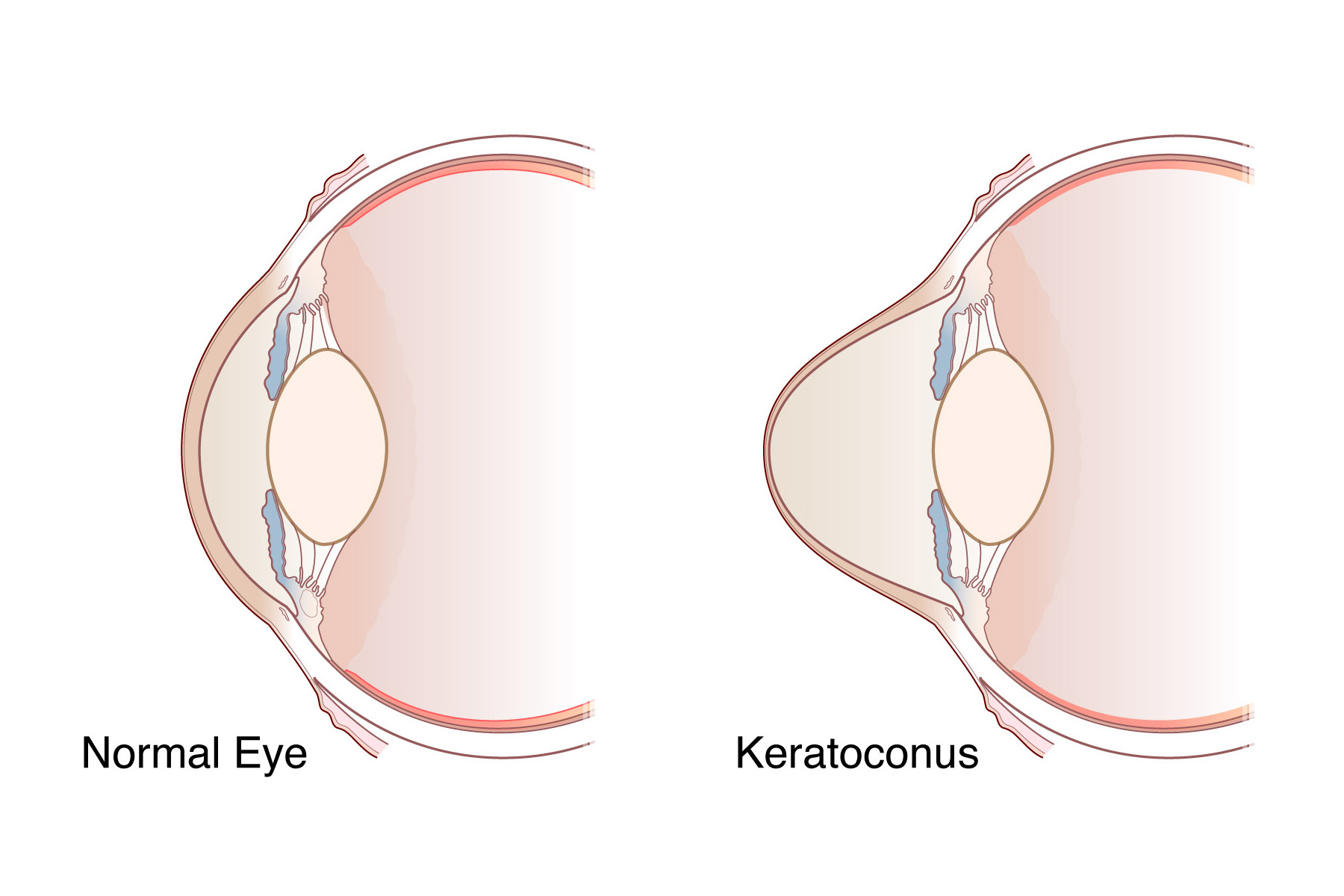
Keratoconus (cone shaped cornea) occurs when the cornea, the window of the eye, begins to bulge forward distorting the surface of the eye and vision. Think of a paper clip, if you bend it over and over it finally reaches a tipping point and breaks. It is similar with eyes, continual rubbing or pushing on the eyes gradually reaches a tipping point where the tissue begins to give way and bulges out. Like a paper clip, it first becomes weakened and then breaks. When the inner surface of the cornea breaks, it lets a lot of fluid into the cornea causing it to swell and this is called hydrops. With hydrops, there is a rapid loss of vision, the cornea looks white, and as it heals it can cause scarring.
A variety of factors contribute to when the breaking point is reached in an individual’s eye and the cornea begins to bulge out causing keratoconus. These factors include the thickness of the cornea, the intensity of the rubbing or pushing on the cornea, age (because the corneal tissue is softer and more pliable in children and gets stiffer with age), family history, and related conditions that cause itching such as allergies and atopic dermatitis. Keratoconus is also more common in conditions like Down’s Syndrome.
SMILE, Lasik, and PRK can make the progression of keratoconus more likely as they remove tissue and make the cornea thinner – that is why we tell all of our Lasik patients to never rub or push on their eyes. It is also why prior to doing these surgeries, screening tests are done to rule out either the predisposition to or the actual finding of keratoconus. It is important that if vision drops with increasing astigmatism or nearsightedness after laser refractive surgery, one should have their eyes checked to rule out ectasia. We have treatments that can stop the progression of the ectasia, but not particularly reverse thinning that has already occurred – therefore early treatment is best.
Keratoconus and corneal ectasia – what are they and what are the treatments
Price Vision Group is one of only a few places in the US to offer a wide range of treatments for keratoconus and related conditions.
What are the treatment options for keratoconus or ectasia following LASIK, SMILE, or PRK?
The treatments for keratoconus are the same as for ectasia after Lasik, SMILE, and PRK.
Price Vision Group offers a variety of treatments for keratoconus and corneal ectasia, depending upon the stage of the disease.
Cornea Crosslinking is a first line treatment
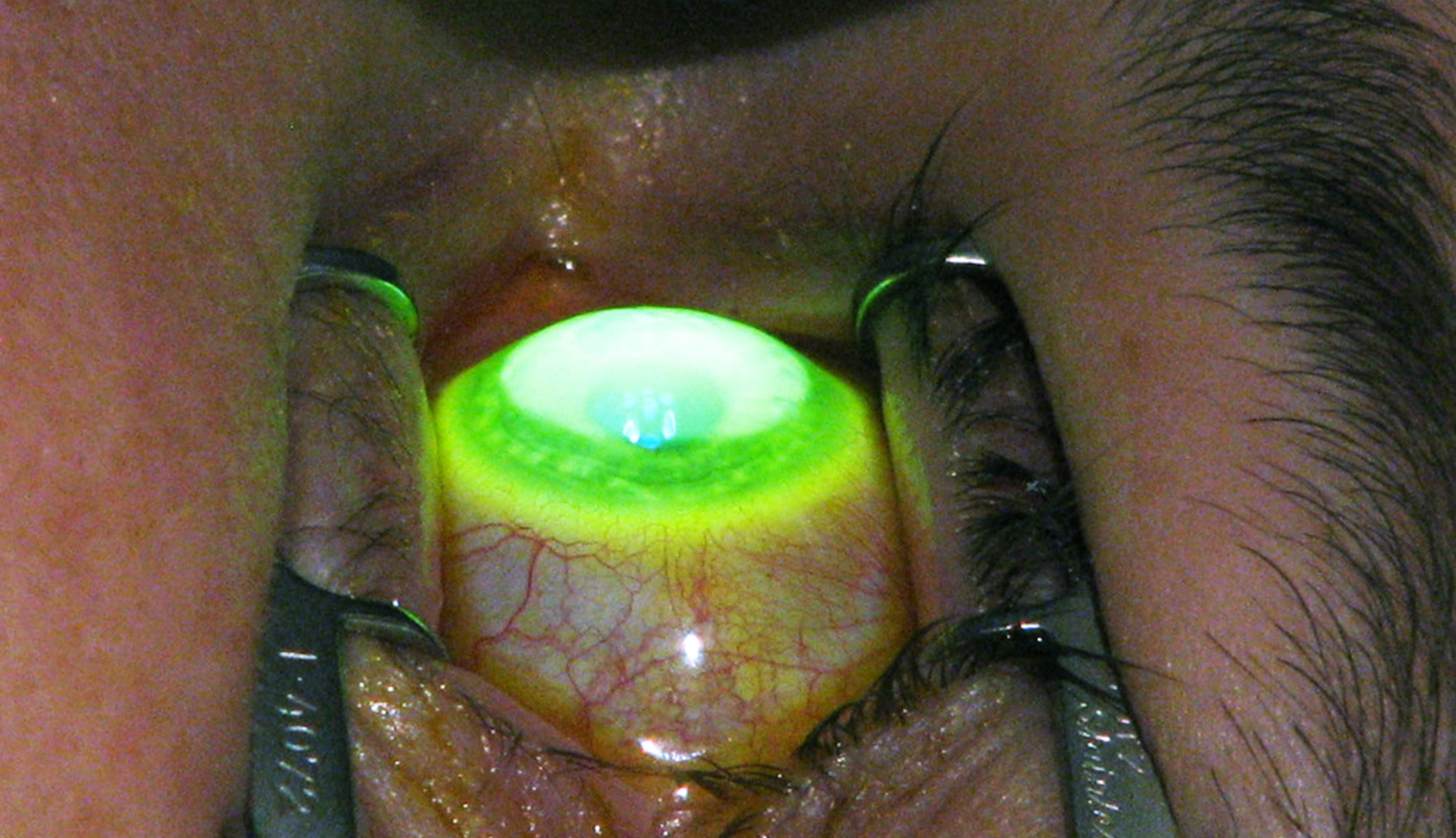
Cornea crosslinking stiffens the cornea, can stop the progression of the corneal bulging, and to some extent can reverse some of the changes. Crosslinking involves soaking the cornea with Vitamin B2 (riboflavin) and then shining an ultraviolet light on the cornea. The riboflavin and the ultraviolet light do not produce changes in the cornea by themselves, but when combined, they produce a photochemical reaction that strengthens the cornea, makes it stiffer, and thereby stops or slows the bulging process.
Crosslinking is facilitated by removing the epithelium (the skin covering of the cornea) to allow the riboflavin to more effectively soak into the corneal tissue. After removing the epithelium, it takes a few days for it to heal over, and until it heals the eye can feel uncomfortable and painful. A variety of techniques can facilitate penetration of the riboflavin through intact epithelium. However, these techniques have not been shown to be as consistent at stopping keratoconus or ectasia progression or to be as effective at reducing corneal bulging as removing the epithelium, suggesting it is the most reliable choice.
Haze in the cornea is common after epithelium-off procedures, and this has often been noted to be sign of the effectiveness of the treatment. The haze usually clears up within 1 to 3 months or sooner.
When should keratoconus be treated?
It’s important to catch and treat keratoconus early to keep it from getting worse! We recommend crosslinking for anyone less than 32 years of age who is exhibiting signs of keratoconus, because the likelihood of progression without treatment is very high in young adults, adolescents and children. Keratoconus can start at a very young age. We have treated children as young as 10 years old. The goal is to stop progression and stabilize the cornea as soon as possible.
We recommend crosslinking to patients over 32 years of age who have significant corneal thinning, recent worsening of vision, and/or recent progression of corneal bulging documented with imaging. As people age, their corneas (like a lot of the parts of the body) get stiffer and the keratoconus can stabilize naturally. Therefore, we require signs of recent progression before treating older patients.
Post-refractive surgery ectasia is very likely to progress at any age once it is diagnosed, so we do not have any age or signs of progression requirement for treating post refractive surgery ectasia.
Early treatment allows individuals with mild keratoconus to continue to use glasses or soft contact lenses for vision correction, and it allows those with moderate keratoconus to use a rigid or hard contact lens for vision correction and avoid more serious surgery, such as a corneal transplant.
A recent study estimated the value of crosslinking in terms of long-term quality of life and reduced future medical costs at $170,000-$250,000. The benefits included better vision, reduced surgical and medical costs by preventing the need for transplants and scleral lenses, and reduced future risk. (Link to the study: https://pubmed.ncbi.nlm.nih.gov/40981503/ )
Contact lenses
- Contact Lenses
- Corneal Transplants
- Anterior Lamellar Keratoplasty with Ring Segments (ALKRS) / Cornea Tissue Addition Keratoplasty (CTAK, CAIRS)
- Deep anterior lamellar keratoplasty (DALK)
- Laser cut DALK
- Full Thickness Penetrating Keratoplasty (PK, PKP) Transplants
- Recurrent Keratoconus after Previous PK / PKP
Contact lens materials and designs such as scleral lenses have greatly improved in the last few decades, and most people with keratoconus can get by with these lenses for improved vision. Keep in mind though that the contact lenses do not stop the continued bulging of the cornea, or progression of the keratoconus – only crosslinking will do that.
Additionally, scleral lenses can lead to hypoxia of the corneal surface when worn too long during the day. As keratoconus progresses and patients become reliant on wearing scleral lenses just to be able to see, it becomes harder to limit wear and patients tend to choose to wear them continuously, jeopardizing the long-term health of their eyes. Early cross-linking treatment to avoid needing rigid contacts is ideal, but scleral lenses are generally still preferable to a cornea transplant.
Penetrating full thickness transplants (PK, PKP), partial thickness deep anterior lamellar transplants (DALK), and tissue addition anterior lamellar transplants (ALKRS, CTAK, CAIRS)
When the keratoconus progresses to the point where vision is not improved enough with contact lenses or someone is unable to successfully wear contact lenses, then corneal transplants become the primary treatment option.
Price Vision Group has published extensively on corneal transplants, and to our knowledge performs more corneal transplants than any other center in the United States. Price Vision Group surgeons are experts in this area and have trained surgeons from around the world. Our doctors routinely give talks on transplants both in the U.S. and internationally.
Most patients with mild or moderate keratoconus can have improved vision with tissue addition surgeries if the central cornea is clear without scarring. Price Vision Group will typically recommend CTAK. The advantages of CTAK or CAIRS (a similar procedure) are that no tissue is removed from the patient’s cornea and nothing is actually transplanted across the central, or visual area of the cornea. A channel is made in the mid portion of the cornea either manually or with a laser. Then a specially shaped portion of donated cornea tissue is inserted into the channel. The insert is custom shaped to the patient’s eye to modify the shape of the patient’s cornea to provide improved vision. The CTAK tissue is processed and treated so there are no viable cells in it. Down time is just a few days at most, and it is done with just topical anesthesia as the cornea can be completely numbed with just eye drops.
We have moved away from using plastic inserts as the tissue addition procedures are more reproducible and now have over 11 years of follow up.
When the cornea is too thin to safely do CTAK or there is central scarring, then we recommend DALK. With DALK we remove all the structural area of the central cornea, the stroma, down to the inner layer called Descemet’s membrane. Leaving Descemet’s membrane and the underlying endothelium greatly reduces the risk of immunologic rejection and allows less use of topical corticosteroids so the graft heals more quickly and securely. The new cornea comes from someone who has died and has donated their cornea. The cornea is fresh as compared to the CTAK corneal grafts, which use preserved tissue. DALK does require sutures to hold the graft in place and the sutures are typically removed 6 to 8 months after surgery. Once sutures are removed then we can determine the degree of visual improvement. About a third of people with DALK continue to wear contact lenses either for treatment of residual irregularities or just so they do not have to wear thick glasses. With DALK, there is always a chance that the recipient’s thin inner corneal membrane may break, which would turn the transplant into a full thickness penetrating keratoplasty (described in more detail below).
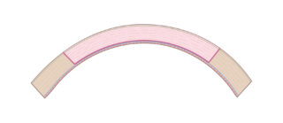
With DALK, the thin layer of bulging recipient tissue that is left in place gets compressed by the donor tissue that is added and this can lead in some cases to some folds in the remaining recipient tissue, which can limit visual recovery. There is always some compression and folding of the tissue, but usually it does not interfere with vision. If it does interfere with vision, then a partial thickness graft can be done where just the inner layer with folding is replaced with a new thin layer of donor tissue and sutures with a long recovery can be avoided. That is a transplant call DMEK.
Corneal transplants are typically performed using a round blade called a trephine, which is like a small cookie cutter. The trephine is used on the patient’s cornea and on a donated cornea and then the donated cornea is sutured onto the patient’s eye – like changing a window pane in a window.
The cornea is made in layers like plywood, and just like if you cut out a circular area of plywood and glue it back, the transplant wound never heals as strong as the original cornea was. The strength of the wound can be enhanced by using a femtosecond laser to make a zig zag (interlocking) incision on the both the patient’s eye and the donor cornea so the two fit together like a tongue-in-grove in carpentry. We offer this option at Price Vision Group for DALK cases. This can be especially helpful with keratoconus, because the donor cornea is often thicker than the patient’s cornea it is being sutured into. The zig zag configuration allows better joining of donor and recipient corneas together than a straight vertical cut does, especially when there is a thickness mismatch.
In cases where there is scarring on Descemet’s membrane, especially if there was previous hydrops, then a DALK can be difficult to impossible to achieve. Then a full thickness transplant is done. As with DALK, sutures are used, but the sutures often remain in place for 12 to 18 months and more corticosteroids are used to prevent immunologic rejection.
Price Vision Group has been actively doing corneal transplants since 1983, and we periodically see patients that we did transplants on 30 or more years ago. For those who had transplants for keratoconus, the recipient or remaining portions of their own cornea sometimes continue to have thinning and bulging outside the grafted area causing astigmatism and distorting the vision. In these cases, if the endothelial layer of the cornea is still functioning and doing well, we can reopen the wound of the transplant in the area of the thinning and resuture it. This maintains the graft which has survived and been tolerated well without rejection for decades. In many cases, the thin areas of the cornea can also be crosslinked to help with healing and minimizing further thinning in the future.
The US Crosslinking Scandal
Currently in the US, there is one approved crosslinking treatment available. This is an epithelial on treatment call EPIOXA and it requires a special set of goggles to wear to apply extra oxygen to the cornea. At the time of writing this, the current cost for a doctor or facility to purchase the supplies to do one treatment is $78,500! Outside the US, crosslinking supplies can be obtained often for less than $100. There are also questions regarding the effectiveness of this epithelial on treatment compared to epithelium off treatments, based on clinical trial data. Also, EPIOXA is not approved for treatment of ectasia after Lasik, SMILE, or PRK. Other investigational crosslinking treatment options, which can be dramatically less expensive, are available, and we offer one at Price Vision Group.
**Eyes can be crosslinked a second time if signs of progression develop, and an epithelium off procedure can be done in eyes with previous epithelial on treatments if there are signs of progression.
Cataracts and Keratoconus
Everyone gets cataracts if they live long enough as they are a natural part of aging where the crystalline lens in the eye becomes cloudy. Keratoconus eyes also develop cataracts and at some point need to have them removed. If the keratoconus is severe and will need a transplant (either PK / PKP or DALK), then the cataract surgery should always be done after the sutures have been removed from the transplant so that the cataract surgery can help mitigate the residual astigmatism, nearsightedness, for hyperopia (farsightedness) after the transplant. If the cataract is done first before the transplant or before suture removal, then a patient may be left with thick glasses in one eye requiring full-time contact lens use to allow the two eyes to work together.
Because keratoconus distorts the surface of the cornea, it also distorts the imaging we use to calculate the appropriate lens power to place in the eye during cataract surgery. With mild cases of keratoconus, doctors can come somewhat close with the testing devices and the use of special toric intraocular lenses that correct astigmatism.
Price Vision Group has developed a two-stage option for cataract surgery for keratoconus eyes to help reduce the need for visual correction afterwards. With the two-stage process, in the initial cataract surgery, we place a standard lens that does not correct astigmatism and is targeted to leave the eye slightly nearsighted so it is easier to see a contact lens and place it in the eye after surgery. Then one to 2 months after surgery the eye is checked to see what the residual glasses prescription is. At that point a patient has 3 options:
1
If the vision is correctable with glasses or they are happy with the result or their uncorrected vision without glasses, they need no further treatment.
2
If a glasses prescription is not helpful and the vision is not acceptable, then the patient could go back into a contact lens for acceptable vision.
3
If the glasses prescription can improve the vision to an acceptable level, but the patient would like to be glasses free for either distance or near vision, then the patient can have a refractive lens exchange where the original intraocular lens is removed and replaced with a new one that corrects the residual astigmatism, nearsightedness, or hyperopia that they had. Note that being glasses-free for both near and distance vision is not an option, because multifocal lenses do not function well in keratoconus eyes. We have shown there are significantly better visual results with this staged technique than placing an astigmatism or toric lens at the time of cataract surgery in keratoconus eyes. (*)
A big advantage of the staged technique is that it avoids placing a toric or astigmatic intraocular lens implant in an eye that could eventually need a contact lens due to irregularities from the keratoconus. If a patient has a toric lens in the eye and then tries to wear a rigid contact lens like a scleral lens, the lens will correct the irregularities from the keratoconus but there will be residual astigmatism from the intraocular lens. Trying to have the contact lens also correct for the astigmatic correction in the intraocular lens can be very difficult.
Potential draw backs to the staged approach include:
- Having to have the second surgery
- The second surgery is a refractive procedure and not covered by insurance and therefore is cash pay for the patient.
- If there are problems with the cataract surgery, or when the going back to do the second surgery such as week support of the capsule to place the lens implant in, then the second surgery would not be possible to either do or to finish.
* Goebel GJ, Price DA, Price MO, Price FW. Functional approach to IOL selection in eyes with combined cataract and keratoconus with an option for refractive lens exchange. J Refract Surg 2024;40(4):e208-217.
What if you do not have insurance coverage or have a high-deductible health plan?
Price Vision Group typically does surgery in a free-standing surgery center or in an office-based surgery suite, and both of these options are much less expensive than hospital based or hospital associated surgeries. We treat many patients without insurance coverage from across the state of Indiana, the US, and internationally. If interested, our staff can help you understand the costs and potential financing for the procedures that you need.
References on DALK
- Saint-Jean A, Soper M, Den Beste K, Iverson S, Price MO, Price FW. Technique for Ensuring Type I Bubble Formation for Pre-Descemet Endothelial Keratoplasty Preparation. 2019;38:1336-1338.
- Wileman JM, Price MO, Price FW Jr. Identification and management of a central type 2 big bubble in deep anterior lamellar keratoplasty. EBCT 2023;2:e0018.
- Gonzalez A, Price MO, Feng MT, Lee C, Arbelaez JG, Price FW Jr. Immunologic Rejection Episodes After Deep Anterior Lamellar Keratoplasty: Incidence and Risk Factors. Cornea 2017;36:1076-1082.
- Burkhart ZN, Feng MD, Price MO, Price, FW. Handheld Slit Beam Techniques to Facilitate DMEK and DALK. Cornea 2013;32:722-4.
- Price MO, Price FW. Deep anterior lamellar keratoplasty: coming of age. Br J Ophthalmol 2010;94:1275-6
- Price FW, Price MO, Grandin JC, Kwon R. Deep anterior lamellar keratoplasty with femtosecond-laser zigzag incisions. Journal of Cataract and Refractive Surgery 2009;35:804-808.
- Price FW. “Small bubble technique” helps “big bubble technique”. British Journal of Ophthalmology, 2007; 91:1260-1.
- Price FW Jr. Intraoperative optical coherence tomography: game-changing technology. 2021;40:675-678.
Keratoconus Detection
- Lopes BT, Ramos IC, Salomao MQ, Guerra FP, Schallhorn SC, Schallhorn JM, Vinviguerra R, Vinciguerra P, Price FW Jr, Price MO, Reinstein DZ, Archer TJ, Belin MW, Machado AP, Ambrosio R Jr. Enhanced tomographic assessment to detect corneal ectasia based on artificial intelligence. Am J Ophthalmol 2018;195:223-232.
- Tenkman L, Price MO, Price FW. Keratoconus onset after age 50. J Refract Surg 2012; 28:436-8
- Guerra FP, Price MO, Price FW. Is central pachymetry asymmetry between eyes an independent risk factor for ectasia after LASIK? J Cataract Refract Surg 2010;11:2016-7.
- Wileman JM, Price MO, Price FW Jr. Case of Progressive Keratoconus With Newly Diagnosed Pellucid Marginal Degeneration After Corneal Cross-Linking. Cornea 2024;43:257-260.
Crosslinking and Keratoconus
- Lautert J, Doshi D, Price FW Jr, Price MO. Corneal Epithelial Remodeling After Standard Epithelium-off Corneal Cross-linking in Keratoconic Eyes. J Refract Surg. 2018;34:408-412.
- Price MO, Feng MT, Price FW Jr. Patient satisfaction with epithelium-off corneal crosslinking. J Cataract Refract Surg 2018;44:323-328.
- Price MO, Fairchild K, Feng MT, Price FW Jr. Prospective Randomized Trial of Corneal Cross-linking Riboflavin Dosing Frequencies for Treatment of Keratoconus and Corneal Ectasia. 2018;125:505-511.
Other Crosslinking
- Price MO, Price FW Jr. Corneal cross-linking in the treatment of corneal ulcers. Curr Opin Ophthalmol 2016;27:250-5.
- Price MO, Tenkman LR, Schrier A, Fairchild KM, Trokel SL, Price FW. Photoactivated Riboflavin Treatment of Infectious Keratitis Using Collagen Cross-linking Technology. J Refract Surg 2012;28:706-13.
- Price MO, Price FW Jr. Corneal cross-linking in the treatment of corneal ulcers. Curr Opin Ophthalmol 2016;27:250-5.
- Price FW Jr, Tefasse Z, Frances KD, Feng MT, Gang A, Price MO. Assessment of Corneal Crosslinking for the Treatment of Corneal Neovascularization With and Without Associated Infection. Cornea. 2025 Apr 2. doi:
General PK survival rates and risk factors includes KC
- Thompson RW, Choi DM, Price MO, Price FW. Noncontact optical coherence tomography for measurement of corneal flap and residual stromal bed thickness after laser in situ keratomileusis. J Refractive Surgery, 2003, 19:507-515.
- Price MO, Thompson RW, Price FW. Risk factors for various causes of failure in initial corneal grafts. Archives of Ophthalmology, 2003;121:1087-1092.
- Cornea 2000 Nov;19(6):813-816.
- Loden JC, Price FW Jr. Price graft-over-host technique to manage positive pressure during penetrating keratoplasty. J Cataract Refract Surg. 1998;24:736-8.
- Price FW, Whitson WE. Graft survival in four common groups of patients undergoing penetrating keratoplasty. Ophthalmology 1991;98:322‑328.